The effects of a commonly used anesthetic drug on memory and thinking appear to be temporary in young and old patients, according to a new study in animals.
The research, published in this week's issue of the journal PLOS ONE, was conducted by biologists at Weill Cornell Medical College and the University of California, San Diego in response to concerns, arising from multiple studies on humans over the past decade, that exposing children, as well as elderly adults, to general anesthetics may increase their susceptibility to long-term cognitive and behavioral deficits, such as learning disabilities.
"There is concern now about cognitive dysfunction from surgery and anesthesia--how much these effects are either permanent or slowly reversible is very controversial," says Dr. Hugh Hemmings, Jr. , chair of anesthesiology at Weill Cornell and one of the two senior authors. "It is not clear whether the residual effects after an operation are due to the surgery itself, or the hospitalization and attendant trauma, medications and stress--or a combination of these issues."
He added that another factor that may lead to some of the delayed or persistent cognitive effects after surgery is brain inflammation resulting from the operation.
Dr. Shelley Halpain, a professor of biology at UC San Diego and the Sanford Consortium for Regenerative Medicine, who co-headed the investigation, says that "because the clinical studies haven't been completed, preclinical studies, such as ours, are needed to define the effects of various anesthetics on brain structure and function."
The team of biologists examined one of the most commonly used general anesthetics, a derivative of ether called isoflurane that is used to maintain anesthesia during surgery.
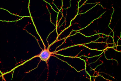
In the experiments at UC San Diego headed by Dr. Jimcy Platholi , a postdoctoral researcher in Halpain's lab who is now at an instructor of pharmacology in anesthesiology at Weill Cornell, the scientists used neurons from embryonic rats taken from the hippocampus, a part of the mammalian forebrain essential for encoding newly acquired memories and ensuring that short-term memories are converted into long-term memories. The researchers cultured these brain cells in a laboratory dish for three weeks, allowing the neurons time to mature and to develop a dense network of synaptic connections and "dendritic spines"—specialized structures that protrude from the dendrites and are essential mediators of activity throughout neural networks.
"Evidence from animal studies indicates that new dendritic spines emerge and existing spines expand in size during learning and memory," Dr. Halpain said. "Therefore, the overall numbers and size of dendritic spines can profoundly impact the strength of neural networks."
Using neurons in culture, rather than intact animal brains, allowed the biologists to take images of the synapses at high spatial resolution using a variety of advanced techniques, an effort led by Dr. Karl Herold, a research associate in the Hemmings laboratory and a co-author of the study.
The researchers investigated whether brief exposure to isoflurane would alter the numbers and size of dendritic spines, so they applied the anesthetic to the cultured rat cells at concentrations and durations (up to 60 minutes) that are frequently used during surgery.
"We clearly see an effect--a very marked effect on the dendritic spines--from use of this drug that was reversible, suggesting that it is not a toxic effect, but something more relevant to the pharmacological actions of the drug," Dr. Hemmings says."Connecting what we found to the cognitive effects of isoflurane will require much more detailed analysis."
The study was supported by grants from the National Institutes of Health (MH087823 and GM58055).