Fleeting blindness, debilitating migraines and a constant pounding sound had all become part of everyday life for Allyson Tlacoxolal. But when the New Jersey mom couldn't see for more than 30 seconds at a time and her vision didn't immediately return after she blinked a few times, Tlacoxolal became truly terrified.
 and Dr. Athos Patsalides (right). Tlacoxolal participated in a clinical trial with the Weill Cornell Brain and Spine Center to treat symptoms caused by her pseudo")
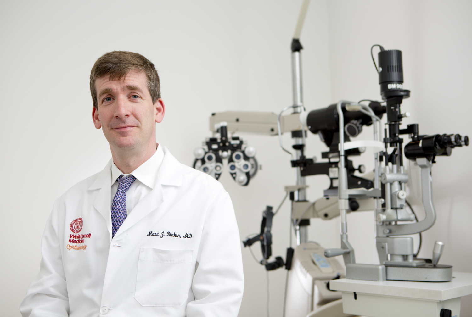
Allyson Tlacoxolal, 33, at her three-month follow-up appointment with Dr. Marc Dinkin (left) and Dr. Athos Patsalides (right). Tlacoxolal participated in a clinical trial with the Weill Cornell Brain and Spine Center to treat symptoms caused by her pseudotumor cerebri condition. Credit: Weill Cornell Brain and Spine Center.
"I always just thought it was because of migraines, because you hear that it can cause blurred vision," said Tlacoxolal, 33. "A couple of times a day I'd see white and have to continually blink until my vision returned. I called these 'blind episodes.' It was very scary and would happen sometimes when I was driving too. That day I just got so scared to the point where I was like, 'Am I never going to see again? Am I not going to be able to blink my vision back in?'"
Tlacoxolal had already been diagnosed with a pseudotumor cerebri, a condition caused by unexplained pressure inside the skull. While it is not actually a brain tumor, a pseudotumor mimics the symptoms of one. The increased pressure can cause swelling of the optic nerve and vision loss, explaining Tlacoxolal’s frequent episodes of blindness.
At the time, Tlacoxolal's doctors recommended placing a shunt in her brain to drain excess cerebrospinal fluid, which would hopefully decrease the pressure on her skull and her symptoms. She put off surgery for another five years, but decided to take action after learning that her spinal fluid pressure was extremely high. She transferred to NewYork-Presbyterian/Weill Cornell Medical Center from central New Jersey for the shunt surgery. As she prepared herself mentally for the procedure, she was surprised to learn that she might not need it after all.
"While Allyson was in the hospital, we did a special MRI study and recognized a narrowing of the large veins in her brain," said Dr. Athos Patsalides, an assistant professor at Weill Cornell Medical College who, along with Dr. Marc Dinkin, is running a clinical trial for patients like Tlacoxolal. "Even though she was scheduled for the shunt, the surgeon in charge called me to let me know she could be a good candidate for the venous stent trial."
Instead of placing a shunt in her brain to drain excess fluid, Dr. Patsalides and Dr. Dinkin proposed inserting a stent into the narrowed veins in her brain. The stent would permanently prop open the veins and prevent a bottleneck, eliminating the backflow of blood into the brain.
"There are two parts to the procedure," Dr. Patsalides said. "In the first part, called venogram, the patient is awake in the operating room and we make a small incision in their groin. We insert a soft, plastic tube into their veins, snake it up through their neck and into the large veins of the brain to assess whether the narrowing shown in the MRI and CT scans is truly significant and is something that explains the patient's symptoms. The patients have to be awake during the first part for the pressure measurements to be accurate."
The next step is inserting the stent. The procedure requires general anesthesia and medications, like aspirin, to prevent blood clots from forming at the site of the stent. According to Dr. Patsalides, patients may opt to wait a few days before the procedure if their symptoms are not severe. However, Tlacoxolal’s symptoms had progressed to the point that waiting was not an option. As soon as the first test confirmed that she was a good candidate, Tlacoxolal immediately underwent the procedure.
"It was not easy to be part of a clinical trial and it was a very scary thing at first … but I can’t tell you enough now how amazing it is,” Tlacoxolal said three months after undergoing the procedure. “I've been feeling completely wonderful, my headaches and the noises in my ears are gone, like from the minute I woke up from surgery. I haven't had any blind episodes at all and am completely headache-free."
Both Dr. Patsalides and Dr. Dinkin presented preliminary findings from the clinical trial this month at annual meetings of the Association for Research in Vision and Ophthalmology and the American Society of Neuroradiology. The trial is still enrolling patients. After the surgery, patients should expect follow-up appointments one, three and six months after the procedure, and an additional appointment every six months afterwards.
For more details on the study, including eligibility criteria, see the listing on http://clinicaltrials.gov/ct2/show/NCT01407809?term=patsalides&rank=1 or contact Dr. Patsalides at 212-746-2821 or atp9002@med.cornell.edu or Dr. Dinkin at 646-962-4297 or mjd2004@med.cornell.edu.