
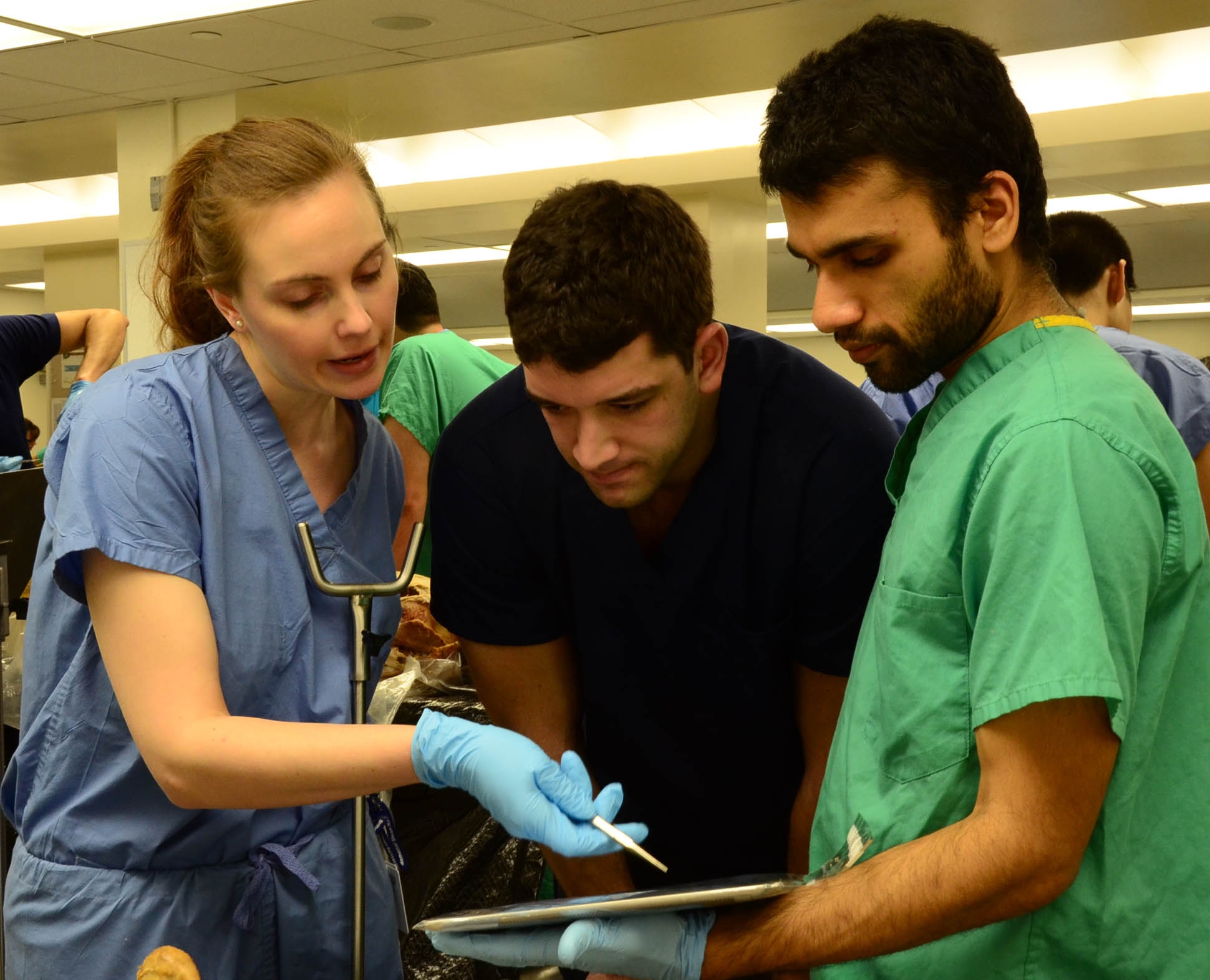
Huddled at the corner of a metal dissection table, four Weill Cornell Medical College first-year medical students survey a cadaver — their first teacher in the complexity and beauty of the human form — with deliberation and reverence.
While Eda Dou, William Cope and Stephanie Sansone stand at its foot, inspecting the bones, muscles and ligaments that together empower human movement and stability, Rivkah Blutstein calls out a list of instructions: how the group should dissect the body, step-by-step.
If it were just a year earlier, Blutstein would be thumbing through the pages of a thick, clunky textbook to guide the dissection. Instead, she was clutching an iPad 2, its weight not even a pound and a half, swiping across the screen to view animated 3-D modeling of the cadaver's foot juxtaposed by photographs of the human structure.
"The pictures are so vivid on the iPad compared to the textbook," marveled Sansone, now a rising second-year student at Weill Cornell.
Less than two years after transforming the medical student learning model from printed course notes and texts to iPads as part of curriculum reform, Weill Cornell has expanded its digital initiative to gross anatomy by purchasing 27 iPads — one for each of the 26 dissection tables stationed in the lab and one for the faculty member running the day's session.
The first-year medical students in the Class of 2016 piloted the technology in the Human Structure and Function class that concluded in the spring. Students and faculty alike say the technology transformed their learning and understanding of human structure and function.
"The use of iPads in lab had a positive impact on student preparedness for the anatomy laboratory sessions, attendance and time spent in the dissection lab," said Dr. Estomih Mtui, director of the gross anatomy and body visualization program at Weill Cornell. "In addition, the information available on iPads also helped students think about the integration of clinical scenarios with the structure and function of each organ that they dissect."
After he began using iPads in the lab, grades for the first exam rose by eight points from the previous year, Dr. Mtui said. For the second exam, Dr. Mtui saw the highest average since 1999, when he started directing the course. While he can't fully credit the improved scores to the iPads, he strongly believes they're at least a contributing factor.
So do his students.
Weill Cornell medical students Jordan Roberts, left, Matt Cohn, center, and Kartik Viswanathan use an iPad to guide a dissection of the human foot during a gross anatomy class in April.
"Everything is centralized, from lecture slides to personalized lecture guides, on one device that's easy and accessible," said Matt Cohn, who was a course representative for the class. "We then used those resources to study in our own rooms."
Gross anatomy, one of the core classes all first-year medical students must take, uses dissection to understand the human body. For the Human Structure and Function class at Weill Cornell, the first-year class is divided into 26 teams of four. In the past, students would need to bring their anatomy textbooks, dissection guides, printed lecture slides and course notes to the lab to guide their dissections.
But that changed in January, when Dr. Mtui introduced iPads to enhance his instruction.
The iPad now houses and safeguards critical resources that were once on paper. Dr. Mtui and his team designed an interactive multimedia program that allowed students to see animated 3-D models of bones, organs and systems. On the iPad, students can manipulate human anatomy, rotating, enlarging or shrinking it, rather than rely on flat, cartoon illustrations on a sheet of paper. Customized, step-by-step directions derived in-house and specifically tailored to Weill Cornell supplant the broad, generic dissection instructions common to wide-release textbooks.
"This way I can customize my curriculum with what we are doing in lab," Dr. Mtui said. "Before that I couldn't. I had to follow the standard instructions found in the textbook. Now I have the control to give students specific instructions that will work for the number of hours available for teaching anatomy."
When put into practice, students found the dissections to be straightforward and effective.
"It's easy for us to locate what they are asking for, rather than guess or wait for a free professor to walk by you," said Noreen Shaikh, 22, president of the Class of 2016.
And at the end of the day, students could take home the materials stored on their iPads as study guides.
Weill Cornell's gross anatomy program joins a small but growing list of medical schools using iPads for dissection, among them the University of California, Irvine, University of Pennsylvania, Harvard and Columbia University College of Physicians and Surgeons.
"The use of iPads in the lab puts Weill Cornell on the leading edge of modern technology in the field of anatomy," Dr. Mtui said.