
It's not an ordinary day on the 13th floor of the Weill Greenberg Center. Weill Cornell scientists are studying an MRI of an adolescent brain, but in a way that defines cutting-edge technology.
Angles and high-definition views of the brain surround the researchers. Images are changed with the flick of the wrist. Components of the image are layered on, separated and even removed from the screen completely to show deep portions of the brain. With a click, the image is enlarged until the scientists are literally standing inside a virtual image of the brain. It is an unparalleled use of technology that has piqued the interest of several other medical schools who are considering investing in similar technology.

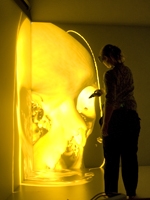
A participant examines the dynamic changes of a 3-D molecular modeling system with a transporter protein immersed in a lipid bilayer environment.
With imaging sciences now playing such a critical role in all areas of medicine — especially diagnostics — Weill Cornell Medical College has spared no expense in outfitting its facilities with the very best imaging technology and software to ultimately benefit both patients and students.
This investment in technology has led to the establishment of the Medical College's new 3-D Immersive Visualization room, part of the Cofrin Center for Biomedical Information in the HRH Prince Alwaleed Bin Talal Bin Abdulaziz Alsaud Institute for Computational Biomedicine at Weill Cornell.
Recently, Dr. Harel Weinstein, the Maxwell M. Upson Professor of Physiology and Biophysics and chairman of the Department of Physiology and Biophysics, who is the director of the Institute, led a demonstration of these stunning capabilities for a delegation visiting from Ontario, Canada.
With the aid of 3-D glasses and a remote control device called a "wand," members of the delegation — which included Carl Zehr, the mayor of Kitchener, Ontario, and his wife, Sandy, along with executives from Christie (manufacturer of the immersion visualization system) — were able to explore the MRI image of the brain with a clarity and precision that was previously unattainable. The images were constructed from data provided by Dr. Barry Kosofsky, professor of pediatrics and chief of the Division of Pediatric Neurology at NewYork-Presbyterian/Weill Cornell. The vivid images were twisted and turned and expanded for close inspection to such dimensions that the visitors were engulfed in color and detailed views of the brain.
At the new 3-D Immersive Visualization room, users are able to delve deep into portions of the human anatomy, including the skull and brain.
"The use of this powerful new tool will increase quickly in all aspects of biomedical research in the College, and will allow us to attract the best and brightest minds in the world," said Dr. Weinstein. "It will provide unique support for Weill Cornell's mission of medical education, clinical excellence and scientific research."
The Weill Cornell Visualization Facility is powered by Christie Mirage HD3 digital projectors, which feature high-definition resolution and projection technology, delivering superior images for molecular modeling, medical diagnosis and scientific research.
Photography by Amelia Panico.