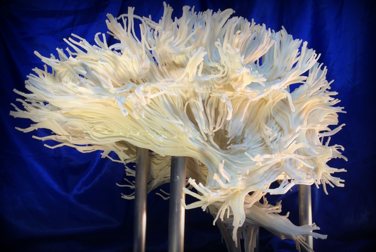
The 2.5-foot brain is the centerpiece of a new exhibit at Philadelphia's Franklin Institute. The exhibit, "Your Brain," provides visitors with the opportunity to explore the brain in themed sections. Dr. Voss' 3-D structure doubles as a work of art and a portrayal of how scientists visualize the pathways that carry out different brain functions.
For Dr. Voss, there was no better time to collaborate with the science center to bring his work to the public. Advanced technology has allowed neuroscientists — and now, the public — to see the brain in ways they couldn't before.
"With the scientific tools that we have now, human neuroscience will have unforeseeable benefits for medicine and humanity in general," said Dr. Voss, the Nancy M. and Samuel C. Fleming Research Scholar in Intercampus Collaborations and associate professor of physics in radiology.
"I think this is what the Franklin Institute wants to convey with their exhibition and I hope that this piece will do its part to make young people interested in the brain as an object of scientific inquiry."
The piece portrays the white matter of the brain, material composed of axons, the long tails of neurons, which facilitate communication throughout the brain. (Its gray matter, which the replica doesn't show, is made up of cell bodies that carry out the body's functions by transmitting electrical and chemical signals.)
Dr. Voss published a similar 3-D image of a patient's white matter in a 2009 review article that later landed in the book "Portraits of the Mind: Visualizing the Brain from Antiquity to the 21st Century." Back in Philadelphia, developers of "Your Brain" stumbled across the image in the book and immediately wanted to bring it to life in the exhibit, which opened last month.
But accomplishing that goal was a complex process. Using an MRI machine at the Citigroup Biomedical Imaging Center, a core facility of the college, Dr. Voss ran a special program called a diffusion tensor MRI scan to capture thousands of images of a consenting person's brain. An algorithm used the images to determine the direction that water travels inside the brain, a property called a diffusion tensor. Another algorithm grouped matching diffusion tensors together to yield a map of the axon bundles that compose the brain's white matter.
The white matter map that emerged created a large 3-D image in which the axon bundles are represented by hundreds of tubes, posing challenges throughout the printing process. After being turned away by 3-D printing companies for nearly a year, the exhibit's project manager, Donna Claiborne, found Direct Dimensions, a 3-D printing company that signed on to the project. Still, in order to make it printable, Dr. Voss had to find a way to reduce the image to about one-fifth of its original size without losing too much of its detail. After he pared it down, Direct Dimensions printed the structure in eight pieces, assembled it, and sent it to the Franklin Institute.
Despite the complicated production process, the piece was well worth the wait, helping to draw more than 47,000 patrons to the exhibit since its debut, Claiborne said.
"It's absolutely beautiful, and it's amazing how much attention it's gotten from all of the visitors," she said. "It's been a great success."